Detecting structure and conformation of single DNA and RNA molecules by Escape-Time Electrometry
Fundamental functions of biological macromolecules, such as proteins and nucleic acids, in all living organisms strongly depend on the molecules’ 3D structures, which are of great scientific interest. However, currently existing methods are bulk measurements, and further require high concentrations. We recently reported a measurement principle – escape-time electrometry (ETe) [1] – that offers a new approach to measuring the effective electrical charge, qm of a single molecule in solution, in real time. Since the effective charge of a molecule in solution is a strong function of its 3D conformation [2], we have demonstrated the ability to directly link a high precision measurement of electrical charge with the 3D conformation of a charged molecule. The ETe approach exploits the spatial confinement of single molecules in an electrostatic fluidic trap. The trap is based on the spatial modulation of the electrostatic interaction energy created in a topographically-tailored fluidic nanoslit [3]. Measurement of the average escape time of single molecules from a trap of known depth allows us to determine the effective electrical charge of a molecular species with high precision.
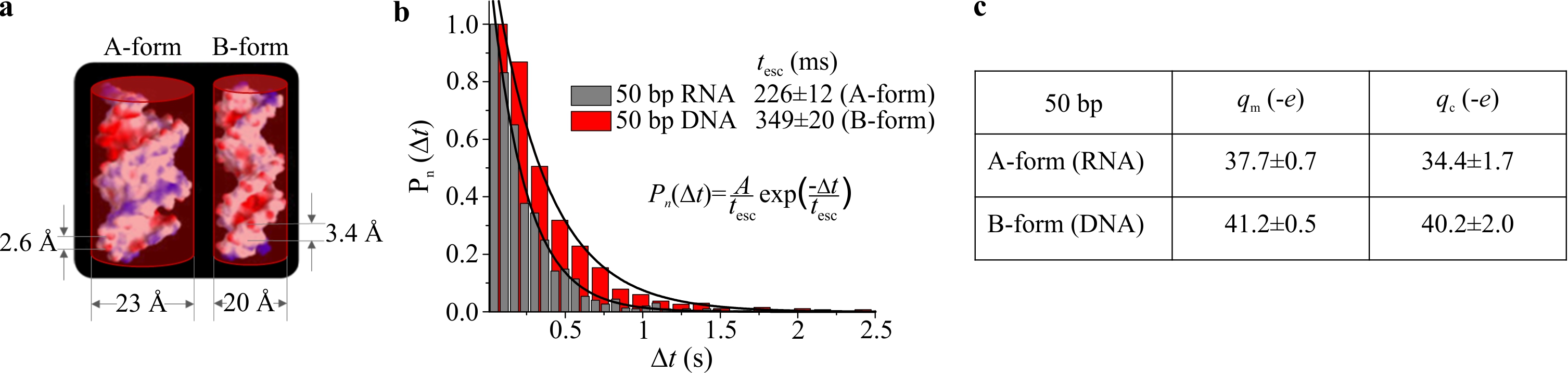
We have performed ETe on double-stranded (ds) nucleic acids with identical base sequences, but different helical structures: A-form (dsRNA) and B-form (dsDNA). Modelling these molecules as uniformly-charged cylinders of different dimensions we also determine theoretically expected values for the effective charge, qc (Fig. a, c). Our measured values of effective charge are in good agreement with the theoretical predictions, and uniquely permit the detection of important structural features of nucleic acids at the level of the single molecule (Fig. b, c).
Furthermore, we are currently performing measurements on single-stranded (ss) RNA and DNA molecules of effectively identical sequence. In contrast to ssDNA, ssRNA is well known to form stable and varied secondary structures, which are in fact critical to biological function. While our preliminary measurements yield a single escape timescale for ssDNA, interestingly they reveal a spectrum of timescales for the equivalent ssRNA species, indicating a variety of folded states. Our results illustrate that ETe will likely enjoy broad relevance in 3D conformation determination in biology.
[1] F. Ruggeri, F. Zosel, N. Mutter, M. Różycka, M. Wojtas, A. Ożyhar, B. Schuler, and M. Krishnan, Nature Nanotechnology, 2017, advance online publication.
[2] M. Krishnan, Journal of Chemical Physics, to appear.
[3] M. Krishnan, N. Mojarad, P. Kukura, and V. Sandoghdar, Nature, 2010, 467, 692-695.